The post-pandemic world has witnessed a surge in artificial intelligence (AI) transforming industries, and healthcare is no exception. With the potential to revolutionize medical diagnosis and patient care, AI has captured the imagination of clinicians and researchers alike. To delve into the practical realities of this exciting shift, Perceptra Company Limited hosted a timely seminar in July 2023: “From Theory to Practice: Sharing Practical Insights on AI in Clinical Radiology.”
During this insightful event, a panel of experts led by Dr. Warasinee, a Faculty member, Institute of Field Robotics, King Mongkut’s University of Technology Thonburi, and co-founder of Perceptra. Her presentation, titled “Navigating the Waves of AI Technology in Healthcare: Trends, Challenges, and Opportunities” provided crucial guidance on navigating the complexities of this transformative technology.
The worldwide trend in Radiology
The current era is witnessing a substantial increase in medical data, with a notable shift – five years ago, the data doubled every three years. In contrast, now, it doubles every two months, a trend expected to continue exponentially.
Fifteen years ago, a radiologist on a 12-hour shift would review 500 images. Today, radiologists are confronted with a staggering 50,000 images during the same timeframe. This translates to today’s radiologists drowning in a sea of medical images that they can never match, a 100-fold increase from fifteen years ago. Radiologist transformed into a machine.
“The artificial intelligence can bring humanity back to medicine,” stated Dr. Eric Topol.
This statement resonates with the growing integration of AI in healthcare. Over 550 AIs received US-FDA clearance as of July 2022, with more than 70% dedicated to diagnostic radiology, placing radiologists at the forefront of AI evaluation. Dr. Warasinee emphasized their critical role as data scientists for patients, analyzing immense data sets to optimize diagnosis and care.
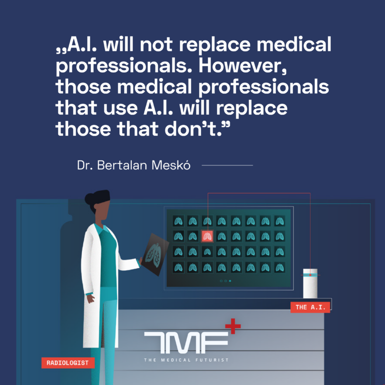
However, many people worry that AI might take their jobs, and it’s normal to feel disheartened about that. But the truth is that several industries, not just radiology, have gone through big changes. For now, people believe that AI will not replace humans, but humans who embrace AI often find that it enhances their capabilities.
Progress in AI for medical images
Understanding the Visionary Power of Deep Nets in Medical Imaging:
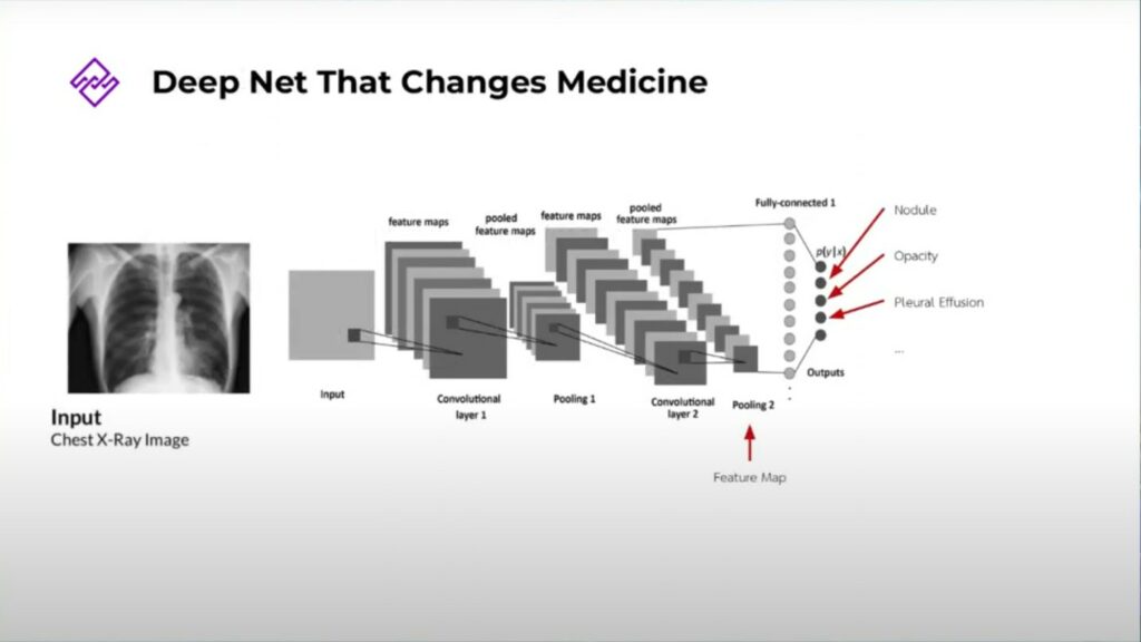
Deep learning, fueled by multi-layered artificial neural networks called Deep Nets, is revolutionizing medical image analysis. Imagine these networks as intricate webs of interconnected nodes, mimicking the structure and information-processing capabilities of the human brain. Just like our eyes perceive complex visuals. This is how AI looks over medical images, translating them into numerical data and extracting meaningful patterns.
Each layer of a Deep Net performs a specific task, progressively building upon the previous one. Imagine zooming in on an image layer by layer. The initial layers might detect basic features like edges and shapes, while deeper layers delve into intricate details. This final layer becomes the master decoder, identifying crucial features like abnormalities within the image.
AI Development Journey
Supervised AI requires a keen understanding of patterns. To achieve this, we partner with radiologists and carefully craft our training recipe: high-quality images, classification labels, and optional localization labels. A million images, seasoned with these ingredients, fuel our AI, teaching it to decipher the secrets hidden within lesions.
The 3 Factors contributing to AI Accuracy
- Labeled data – The AI algorithm requires large quantities of accurately labeled data to learn and improve.
-
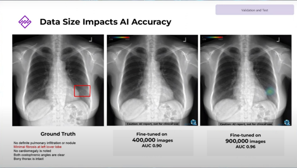
- Quantity matters -Training data size is crucial. Our earlier AI version 1 couldn’t detect fibrosis, while our advanced version can, thanks to a larger and richer dataset.
-
- Quality counts – Accurate labels are essential. To understand the information in the medical report, we focus on identifying the type and location of the lesion. This involves using an AI-driven algorithm to capture keywords and context the radiologist reports.
- Model quality
-
- Architecture suitable for medical image – Convolutional Neural Networks (CNNs) are a common starting point, but medical imaging demands specialized architecture. While the model excels at classifying cats and dogs, classifying X-ray images poses a more specific challenge.
-
- Continuous update – The AI model should have continuous updates through ongoing feedback and updates
- Validation process – AI must be rigorously tested to prove that it meets global medical device standards. Inspectra AI goes through various validation steps, including setting clear inclusion/exclusion criteria, conducting multi-site clinical validation both within and outside the training center, and performing comprehensive software validation.
Current Limitations and Road to Full Adoption
- Limitations:
- Noisy Reports: AI can be misled by unclear or inaccurate medical reports.
- Data Glitches: Image quality issues, foreign objects in scans, and variations in patient positioning can confuse AI.
- Rare Cases and Clinical Context: AI currently struggles with rare medical conditions and situations that require understanding complex medical history.
- Towards Full Adoption:
The 5 Levels of Automation in Medical Procedures
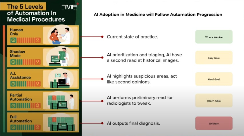
AI is likely to assist in specific tasks, not take over entirely. As you can see in the picture above, 5 levels of Automation in medical procedures. This shows that AI automation may reach partial automation but in Dr.Warasinee’s opinion, this will not go to full automation, at least in our lifetime.
